
Most human proteins remain too small for today’s most powerful biological microscopes to resolve clearly, leaving crucial cellular structures hidden from medical research. A new laser-based innovation developed by UC Berkeley physicists is changing this by upgrading the cryo-EM laser phase plate to reveal biological structures that have long remained beyond reach. This breakthrough is critical for structural biologists and pharmacologists who need to observe how molecules interact in their natural environment.
By dramatically increasing the signal-to-noise ratio, scientists can now observe how molecular machines operate inside living cells. This capability directly impacts drug development, allowing researchers to target diseases at a granular level previously considered impossible. The system adapts phase contrast, a century-old imaging method, for modern cryoelectron microscopy (cryo-EM) and cryoelectron tomography (cryo-ET).
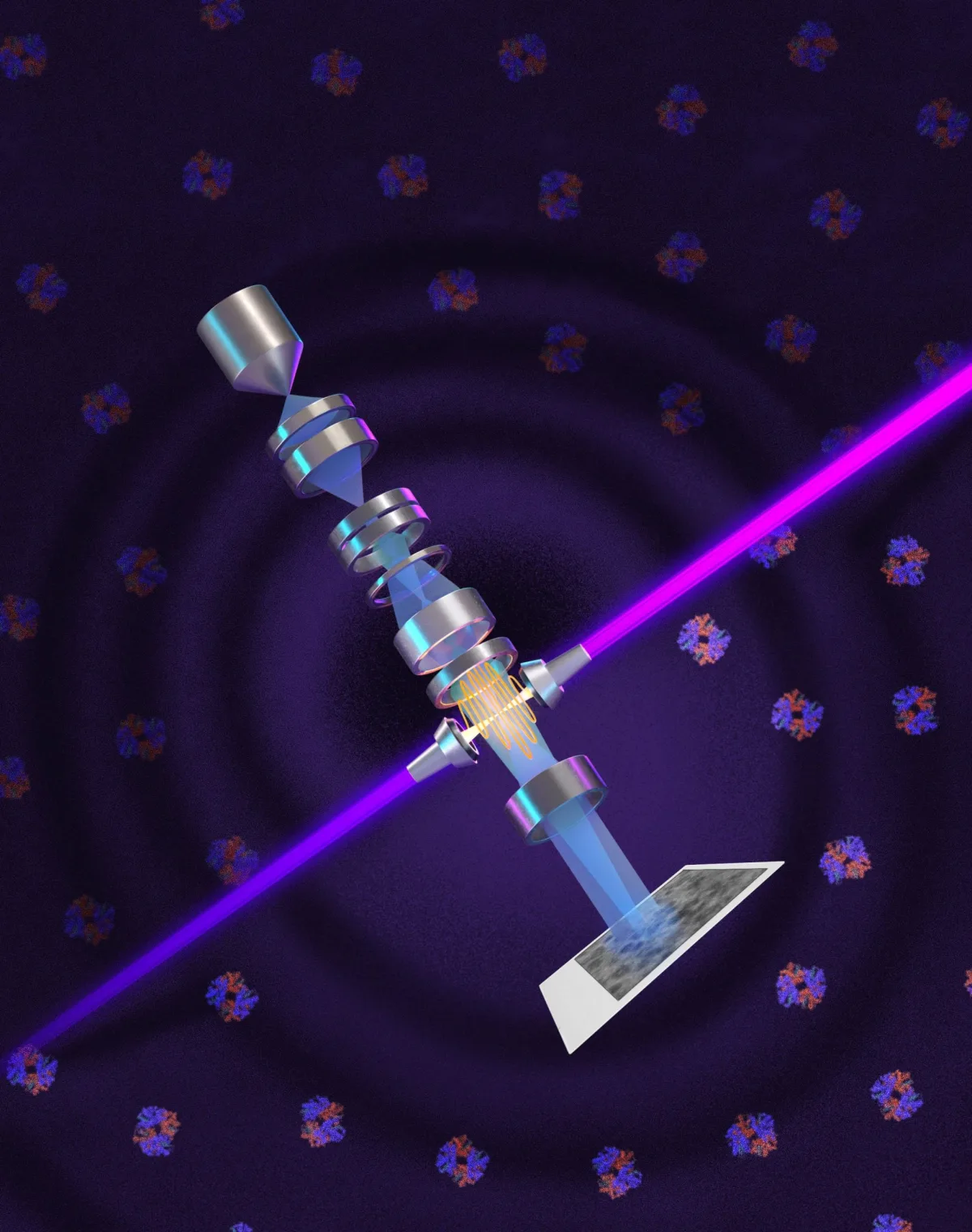
To achieve this, the research team trapped a continuous-wave laser inside a spherical mirrored cavity, reflecting the light over 10,000 times. This concentrates 75 kilowatts of power into a microscopic area, creating an exceptionally intense focus that alters the electron beam's phase without reducing its intensity.
We are going to be able to see how molecular machines operate inside the living cell, in context, for the first time. What was once invisible will become visible - and that changes everything about how we understand disease.
- Stephani Otte, Vice President of Imaging Science, Biohub
Building Theia and Pushing Resolution Limits
The research effort, led by physicist Holger Müller over 15 years, culminated in a customized Thermo Fisher Krios microscope named Theia. Equipped with the new laser phase plate, Theia features extra electron optics that provide superior resolution even before the laser is activated. Biohub, which funded the project, is already developing a second microscope utilizing two perpendicular lasers at lower power to reduce optical distortions.
Current cryo-EM technology struggles to resolve proteins smaller than 70 kilodaltons, a major limitation given that proteins below this size make up roughly 90 percent of the human proteome. During testing on six biological samples, the laser system successfully imaged hemoglobin, a protein near the lower limit of current instruments. Researchers can now image proteins as small as 50 kilodaltons, with future improvements aiming to lower that limit to 17 kilodaltons.
The Proteome Bottleneck Finally Breaks
The introduction of the cryo-EM laser phase plate is not just an optical upgrade; it is a foundational shift for molecular biology. By breaking the 70-kilodalton barrier, this technology unlocks access to the vast majority of the human proteome that has historically been invisible to structural analysis. The ability to clearly distinguish small molecules in the incredibly crowded environment of a cell changes the trajectory of targeted therapeutics.
Furthermore, the sheer power of the 75-kilowatt laser focus demonstrates a remarkable leap in precision engineering. As Biohub develops the next iteration with perpendicular lasers, the potential for cryo-ET to map complex cellular machinery in 3D will likely become the new gold standard for pathology. If the team successfully reaches their 17-kilodalton target, the pharmaceutical industry will gain an unprecedented map of the building blocks of human disease.
This article was written by AI from reporting by scitechdaily.com.