

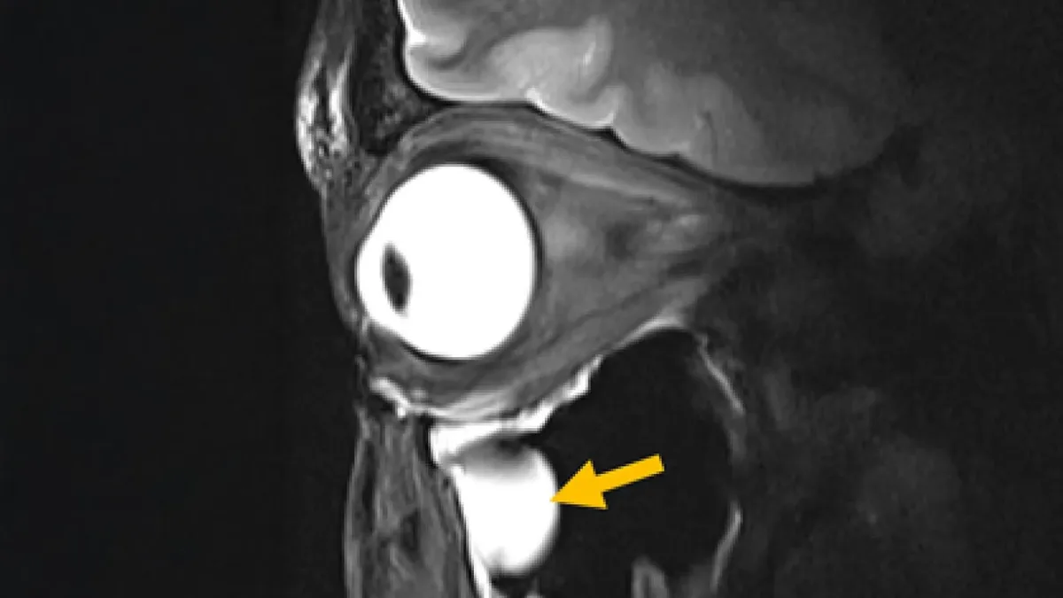
Scientists at the Max Delbrück Center and Rostock University Medical Center have engineered a new MRI metamaterial antenna that dramatically sharpens images of deep brain structures and the human eye. Published in Advanced Materials, the breakthrough redesigns the hardware responsible for transmitting and receiving radiofrequency signals. By integrating advanced engineered materials into existing scanners, the research team has solved a long-standing problem in medical imaging: capturing clear, high-resolution data from anatomically complex regions without extending scan times.
Traditional magnetic resonance imaging relies on antennas, known as radiofrequency coils, to send signals into the body and collect the tissue's response. However, these standard coils often struggle to gather sufficient signal from deep tissues, resulting in blurry images and forcing patients to endure lengthy, uncomfortable scanning sessions. To overcome this, the research team, led by doctoral student Nandita Saha and Professor Thoralf Niendorf, incorporated metamaterials directly into the antenna design.
Metamaterials are engineered structures designed to interact with electromagnetic waves in ways that natural materials cannot. During testing on a 7.0 Tesla MRI scanner, the new antenna successfully guided radiofrequency fields more efficiently. This strengthened the signals from targeted tissues, significantly increasing spatial resolution and accelerating data collection.
It offers the potential to open a window into the eye and into (patho)physiological processes that in the past have been largely inaccessible.
- Professor Oliver Stachs, University Medicine Rostock
The innovation extends far beyond simply taking better pictures of the eye and orbit. Saha explained that their goal was to rethink MRI hardware from the ground up using modern physics. Because the new antenna is compact and lightweight, it can be customized to fit different parts of the body, improving overall patient comfort.
Beyond Diagnostics: Cancer Treatment and Implant Safety
The precision of the metamaterial antenna opens new doors for therapeutic applications. The technology can be adapted to protect sensitive areas of the body during MRI exams by actively reducing unwanted heating around metallic medical implants. This has historically been a major safety hurdle for patients requiring high-field MRI scans.
Furthermore, the researchers noted that the antenna could improve MRI-guided cancer treatments. By directing radiofrequency energy with pinpoint accuracy, the hardware could be utilized for procedures like tumor hyperthermia or thermal tissue ablation, where controlled heat is used to destroy cancer cells.
The research team is already preparing for larger clinical studies involving multiple hospitals. Future iterations of the antenna will be modified to image other vital organs, including the heart and kidneys. Niendorf also confirmed that the design could eventually be adapted for MRI systems operating at magnetic field strengths both lower and higher than 7.0 T, and could even be used to image atoms other than hydrogen, such as sodium and fluorine.
The End of the Million-Dollar Upgrade Cycle
The most disruptive aspect of this breakthrough isn't just the leap in spatial resolution - it is the backward compatibility. Historically, achieving a massive jump in MRI image quality required hospitals to purchase entirely new, multi-million-dollar machines. By proving that metamaterials can be integrated directly into the antennas of existing MRI equipment, this research effectively decouples hardware performance from the core scanner infrastructure.
This approach democratizes ultra-high-field imaging. Facilities that cannot afford to replace their current MRI fleets could simply upgrade their radiofrequency coils to achieve next-generation clarity. As Dr. Ebba Beller noted, innovations in imaging hardware have the potential to transform diagnostics, but the real-world impact will be defined by how quickly this cost-effective upgrade path can clear regulatory hurdles and reach standard clinical practice.
This article was written by AI from reporting by sciencedaily.com.